
The heart attaches to the major vessels and is suspended in the mediastinum by its base, the wide end. The apex describes where the ventricles come to a pointed tip. The anterior region is the ventral aspect of the surface of the heart. The inferior region is the underside of the heart. The posterior region is the backside of the heart.
Fibrous rings provide attachments for valves and muscular fibers and prevent orifices from dilating excessively during ventricular contraction.
Strong fibrous strings called chordae tendineae are attached to the valvular cusps in the ventricles. These strings originate from small mounds of muscle tissue called papillary muscle, which support the chordae tendineae.
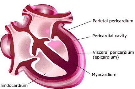
The pericardium is a tough two-layered sac in direct contact with the cardiac surface and separates the heart from other structures in the mediastinum. Fibrous pericardium (protective tough outer layer)- attaches to the bases of the pulmonary artery and aorta, the diaphragm, and sternum. Serous pericardium (thin, delicate membrane)- lines the fibrous sac. The outer layer of this lining is the Parietal pericardium; the inner layer is the Visceral pericardium
Friction between the pericardium and the beating heart is reduced to a minimum by a small amount of lubricating pericardial fluid. Under abnormal conditions, excess fluid may accumulate in the serous pericardial sac developing a pericardial effusion.
Heart Wall
The wall of the heart is composed of three histologically distinct tissue layers-that is, each has a different cellular makeup. The thin outermost layer is the epicardium. The dark red muscle of the heart, the myocardium, is located beneath the epicardium. The myocardium is visible through the epicardium except in places where adipose tissue (fat) has accumulated.
Finally, a thin delicate layer of cells called the endocardium lines the inside surfaces of the myocardium and the valve leaflets within the heart. Although disease may affect any of these tissues, the heart is primarily a muscular organ. In fact, the term myocardium is often used to refer to the entire tissue mass of the heart.
 |
Artwork source:The Heart of Nuclear Cardiology, An Interactive Primer; © 2002 Bristol-Myers Squibb Imaging. Inc.
Chambers
The heart is divided into four distinct chambers with muscular walls of varying thickness. Two thin-walled atria sit atop two thick-walledventricles. The left atrium and right atrium are small chambers located just above the left and right ventricles. The atria are the holding tanks of the heart. The right atrium receives blood from the venous system and the tissues of the body; the left atrium receives oxygenated blood from the lungs. The ventricles are larger as they must propel blood to either the lungs or the tissues of the body.
The heart is divided into four distinct chambers with muscular walls of varying thickness. Two thin-walled atria sit atop two thick-walledventricles. The left atrium and right atrium are small chambers located just above the left and right ventricles. The atria are the holding tanks of the heart. The right atrium receives blood from the venous system and the tissues of the body; the left atrium receives oxygenated blood from the lungs. The ventricles are larger as they must propel blood to either the lungs or the tissues of the body.
A thin muscular wall, the interatrial septum, separates the two atria from one another. A thicker muscular wall, the interventricular septum, divides the ventricles. Separating the atria above and the ventricles below is the atrioventricular septum- the fibrous skeleton of the heart to which are attached not only the atria and ventricles, but also the heart valves and the trunks of the aorta and pulmonary artery. The atria and ventricles are not connected by muscles, only by this dense connective tissue and the valves.
Since the ventricles do the hardest work, their walls are thicker than those of the atria. The muscle fibers of the ventricles are arranged in circular bands, providing the myocardial tissue with the strength and elasticity needed for its continuous pumping activity. The left ventricular wall is approximately three times as thick as the right wall. In order to pump blood through the systemic circulation, the left ventricle must generate three times as much pressure as the right ventricle, which pumps blood only through the low-pressure pulmonary circulation.
No comments:
Post a Comment